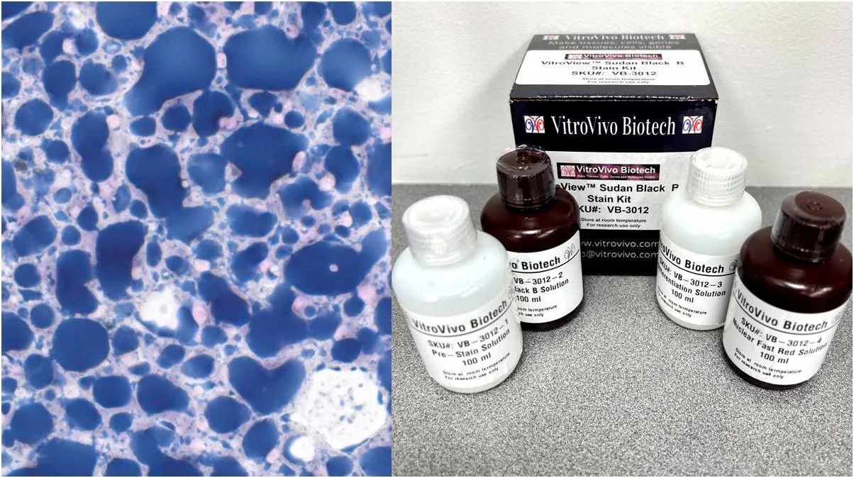
Changes in tissue composition often involve more than one biological process. Fat deposition, extracellular matrix expansion, and structural remodeling can occur in the same experimental model, but they cannot be measured effectively with one stain. A sudan black stain helps researchers visualize lipid-rich material when the goal is to examine fat distribution in preserved frozen tissue.
The broader lesson is that tissue chemistry should guide method selection. Lipids and collagen have different physical properties, respond differently to processing, and require different staining approaches. Treating them as separate endpoints can give researchers a clearer picture of how tissue changes over time.
Start With the Biological Endpoint
Before sectioning begins, researchers should decide whether they are studying intracellular lipid storage, connective-tissue remodeling, or both. That decision affects fixation, embedding, section type, staining chemistry, and even the type of mounting medium used later.
When collagen organization is the focus, a Picro Sirius Red stain can highlight collagen I and III fibers and support evaluation with standard or polarized light microscopy. This makes it useful when researchers need to examine matrix deposition or fiber organization rather than lipid content.
Preserve Lipids Before Staining
Lipid-focused histology requires careful preparation because many routine paraffin-processing solvents remove fats from tissue. A section may retain general morphology while losing much of the material researchers intended to study.
For this reason, Sudan Black B is intended for formalin-fixed frozen sections rather than paraffin-embedded tissue. The stain produces blue-black lipid and fat signals, while nuclear fast red provides contrasting nuclear detail for orientation within the section.
Examine Collagen as a Structural Network
Collagen analysis involves a different question. Researchers may want to determine whether connective tissue has expanded, whether fibers have become denser, or whether matrix remodeling differs between experimental groups.
Picro-Sirius Red produces red collagen staining under ordinary light microscopy. Under polarized light, collagen fibers show birefringence, which can add useful information about thick and thin fiber populations and their organization within the tissue.
Plan Adjacent Sections Strategically
When both lipid and collagen changes are relevant, adjacent sections can support complementary measurements. One frozen section may be reserved for lipid staining, while other suitably prepared sections can be allocated to connective-tissue analysis.
Researchers should record section order and orientation because neighboring sections are similar but not identical. This is especially important when small lesions, deposits, or anatomical regions change quickly across successive cutting levels.
Keep Processing Variables Consistent
Section thickness, fixation time, reagent exposure, washing, differentiation, and mounting can all influence how a stain appears. Standardizing these variables makes comparisons between samples more dependable.
Mounting chemistry also matters. Lipid stains generally require aqueous mounting conditions because organic solvents can interfere with the material being visualized, while collagen protocols may use dehydration and organic clearing before coverslipping.
Use Controls to Catch Technical Problems
Positive control tissue helps confirm that a staining run is functioning as expected. It can reveal problems with reagent performance, staining time, section preparation, or other technical variables before experimental findings are interpreted.
Controls are particularly important in multi-batch studies. Processing them alongside research samples helps investigators distinguish a true change in tissue composition from variation introduced by different staining runs.
Standardize Imaging and Measurements
Microscope settings should remain consistent when images will be compared across experimental groups. Magnification, illumination, exposure, white balance, and polarization settings can all affect the visual appearance of staining.
The analysis plan should also be defined in advance. Researchers may measure lipid-positive area, collagen-positive area, fiber distribution, or region-specific changes, but the same criteria should be applied to every specimen.
Interpret the Two Signals Separately
Lipid accumulation and collagen deposition can occur together, but they represent different biological processes. Increased fat staining does not automatically mean increased fibrosis, and greater collagen deposition does not by itself indicate altered lipid storage.
Treating each endpoint independently first helps prevent overinterpretation. Researchers can then compare the results with morphology, molecular data, treatment groups, or time points to understand how metabolic and structural changes relate within the study.
Conclusion
Tissue remodeling becomes easier to study when researchers select methods according to the material they need to visualize. Lipid-focused and collagen-focused stains provide complementary information, but each requires its own preparation, staining, and evaluation strategy.
Careful specimen handling, appropriate controls, standardized imaging, and clearly defined measurements help turn these stains into reliable research tools. By separating different tissue components analytically, investigators can build a more complete and interpretable view of complex biological change.